Fig. 5.

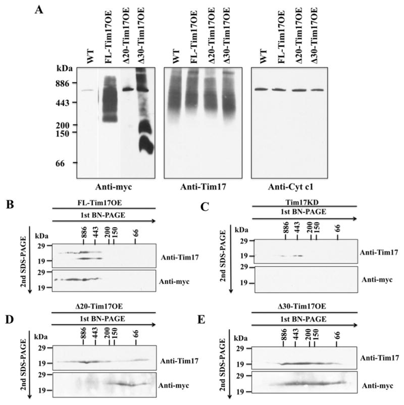
Analysis of the TbTim17 protein complex in mitochondria. (A) BN-PAGE analysis of the mitochondrial membrane protein complexes. Mitochondrial proteins isolated from wild type (WT), FL-Tim17OE, Δ20-Tim17OE, and Δ30-Tim17OE T. brucei grown for 48 h in the presemce of doxycycline were solubilized with digitonin (1%). The solubilized supernatant were clarified by centrifugation at 100,000 × g and analyzed by BN-PAGE. Protein complexes were detected by immunoblot analysis using anti-myc, -Tim17, -cytochrome c1 (Cyt c1) antibodies. Molecular size markers are indicated. (B-E) Gel strips representing the individual lanes for FL-Tim17OE (B), Tim17KD (C), Δ20-Tim17OE (D), and Δ30-Tim17OE (E) samples were excised from the first dimension BN-PAGE and subjected to 12% Tricine-SDS-PAGE. Proteins were transferred to a nitrocellulose membrane, and blots were probed with anti-myc and anti-Tim17 antibodies.
