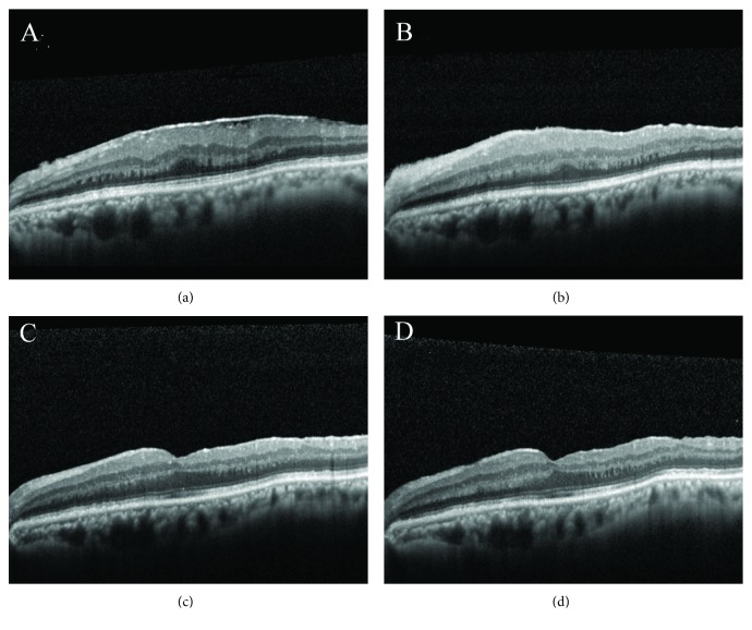
Figure 6.
Pre- and postoperative OCT findings in a patient with ERM receiving vitrectomy, membrane peeling, and maculorrhexis ILM peeling. Preoperative image shows an ERM overlying the macula. The CFT and BCVA were 448 μm and 20/80, respectively (a). The CFT decreased to 425 μm one month postoperatively (b). At six months after operation, the CFT was 306 μm and foveal depression was observed (c). At 12 months after operation, the foveal depression still remained. The CFT further decreased to 289 μm, and the BCVA improved to 20/20 (d).

