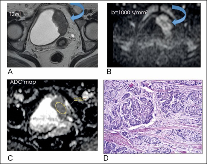
Figure 1.
A 50-years old woman with squamous cell carcinoma, GII, pT3. (A) T2-weighted axial imaging shows a tumor with intermediate signal intensity arising from the left lateral wall of the bladder (curved arrow). (B) Diffusion-weighted image (b = 1000 s/mm2) depicts the tumor as having hyperintense signal intensity with an irregular outline (curved arrow). (C) Apparent diffusion coefficient map: 0.9 x 10⁻³ mm2/s (D) Moderately differentiated Squamous cell carcinoma infiltrating the muscularis propria of the urinary bladder, H&E, x200.

