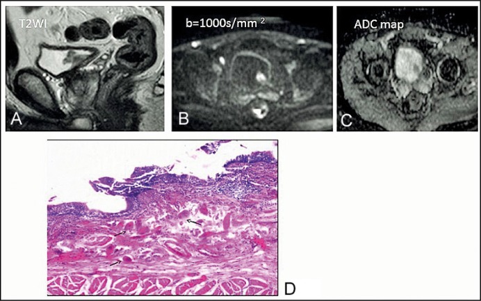
Figure 2.
A 43-year-old man with bilharzial cystitis. (A) T2-weighted imaging shows a tumor with intermediate signal intensity on the left posterolateral side of the bladder. (B) Diffusion-weighted image of the tumor shows restricted diffusion. (C) Apparent diffusion coefficient map: 1.4 x 10⁻³ mm2/s. (D) Bilharzial cystitis showing multiple bilharzial ova (arrows) in the lamina propria of the urinary bladder surrounded by chronic inflammatory cells, H&E, x200.

