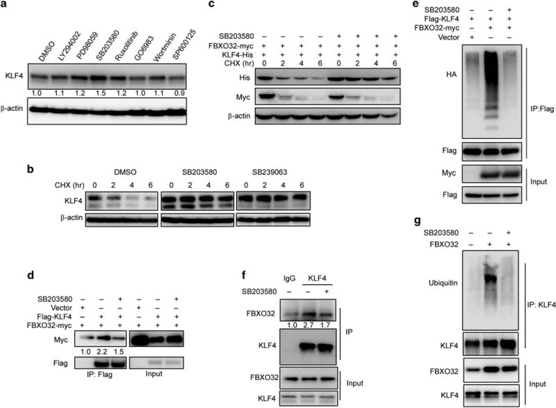
Figure 5.

Active p38/MAPK pathway is necessary for FBXO32-mediated KLF4 degradation. (a) p38 inhibitor leads to KLF4 accumulation. MCF7 cells plated in six-well plate were treated with DMSO or indicated inhibitors for 6 h. The protein level of KLF4 was detected by immunoblot. (b) KLF4 protein half-life is elongated under p38 inhibition. MCF7 cells plated in six-well plate were treated with DMSO or p38 inhibitors (SB203580 and SB239063) for 6 h. Then CHX was added 0, 2, 4 or 6 h before harvest. KLF4 protein level was tested by immunoblotting. (c) p38 inhibitor abolishes FBXO32-mediated KLF4 degradation. HEK293T cells were transfected with His-tagged KLF4 and Myc-tagged FBXO32. Twenty-four hours after transfection, the cells were treated with DMSO or p38 inhibitor for 6 h followed by treatment with CHX for indicated time. Western blot was performed to examine the protein level of KLF4. (d, f) p38 inhibitor disrupts KLF4 and FBXO32 interaction. HEK293T cells were transfected with Flag-tagged KLF4 and Myc-tagged FBXO32 or empty vector. Twenty-four hours after transfection, the cells were treated with DMSO or SB203580 for 6 h followed by 4-h treatment with MG132. The cell lysates were precipitated with anti-Flag M2 affinity gel and the precipitates were analyzed by western blot (d). MCF7 cells cultured in 10 cm dishes were treated with either DMSO or SB203580 for 6 h. The cell lysates were subjected to co-IP analysis by KLF4 antibody. The immunoprecipitates were blotted by FBXO32 antibody (f). (e, g) p38 inhibitor decreased FBXO32-mediated KLF4 ubiquitination. Indicated plasmids were co-transfected into HEK293T cells. Twenty-four hours after transfection, the cells were treated with DMSO or SB203580 for 6 h followed by 6- h treatment with MG132. The cells were subjected to ubiquitination assay and the ubiquitinated KLF4 were detected by immunoblotting with anti-HA antibody (e). FBXO32 overexpressed MCF7 cells cultured in 10 cm dishes were treated with either DMSO or SB203580 for 6 h. The cell lysates were subjected to ubiquitination analysis. Ubiquitinated KLF4 proteins were revealed by immunoblotting with ubiquitin antibody (g).
