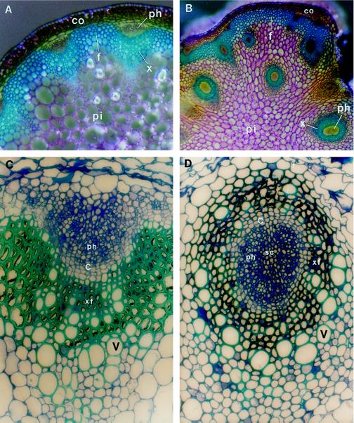
Figure 1.
Vascular patterns in inflorescence stems of the wild type and the avb1 mutant of Arabidopsis. Stem sections of 8-week-old (A and B) or 10-week-old (C and D) plants were stained with toluidine blue. Xylem and fiber cells stained blue because of the presence of lignin in walls. A, Cross-section of the wild-type stem. The individual vascular bundle was collateral, and collectively the vascular bundles were arranged in a ring. B, Cross-section of the avb1 stem. Xylem formed a circle in most bundles, and extravascular bundles were placed in the pith. Arrows indicate the collateral bundles at the periphery of the stele. C, Collateral vascular bundle in the wild type. Xylem was located on the inner side of the bundle, and phloem was located on the outer side of the bundle. One to two layers of cambial initials were evident between xylem and phloem. D, Amphivasal vascular bundle in the avb1 mutant. It was obvious that the phloem was completely surrounded by vessels, a pattern similar to that seen in an amphivasal vascular bundle. In addition, there were one to two layers of cambial initials between xylem and phloem. c, Cambium; co, cortex; f, interfascicular fibers; ph, phloem; pi, pith; sc, sclerified cell; v, vessel element; x, xylem; and xf, xylary fiber. Magnifications, ×102 (A and B) and ×770 (C and D).

