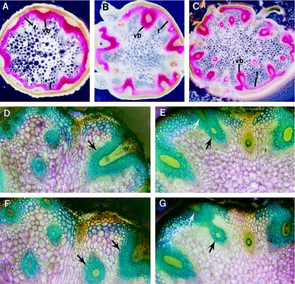
Figure 2.
Abnormal branching and penetration of vascular bundles into the pith of avb1 stems. Serial sections of the internodes were prepared and stained with phloroglucinol-HCl (A–C) or toluidine blue (D–G). It was obvious that the presence of extravascular bundles in the pith was due to abnormal branching and penetration of vascular bundles into the pith (D–G). Arrows point to the branching bundles. A to C, Vascular patterns in stems of the wild type (A), heterozygote (B), and homozygote (C) of the avb1 mutant. D and F, Two adjacent sections of an avb1 internode. E and G, Two adjacent sections of an avb1 internode. f, Interfascicular fibers; vb, vascular bundle. Magnifications, ×52 (A–C) and ×154 (D–G).

