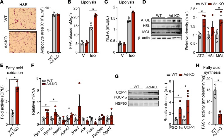
Figure 4. Adipose ANGPTL4 loss enhances lipolysis and oxidative metabolism but reduces endogenous lipogenesis.
(A) Representative H&E-stained sections of white adipose tissue (WAT) isolated from WT mice and in mice with AT-specific knockout of ANGPTL4 (Ad-KO) fed a high-fat diet (HFD) for 4 weeks. Quantification of adipocyte size (right) (n = 3). Scale bars: 200 μm. (B) Ex vivo lipolysis of WAT isolated from WT and Ad-KO mice and treated with vehicle (V) or isoproterenol (Iso) (n = 3). FFA, free fatty acid. (C) In vivo lipolysis in 8-week-old WT and Ad-KO mice (n = 3). NEFA, nonesterified fatty acid. (D) Western blot analysis of indicated proteins in WAT from WT and Ad-KO mice after 4 weeks on HFD (n = 3). Densitometric analysis of the blots is shown on the right panel. (E) Ex vivo fatty acid oxidation of WAT isolated from WT and Ad-KO mice (n = 3). (F) mRNA expression of indicated genes in WAT from WT and Ad-KO mice fed an HFD for 4 weeks (n = 3). (G) Representative Western blot analysis of indicated proteins in WAT from WT and Ad-KO mice fed an HFD for 4 weeks (n = 3). Densitometric analysis of the blots is shown on the right panel. PGC-1α, PPARγ coactivator 1α; UCP-1, uncoupling protein 1. (H) Ex vivo fatty acid synthase (FASN) activity was measured in isolated WAT of WT and Ad-KO mice (n = 3). All data represent the mean ± SEM. *P ≤ 0.05 comparing Ad-KO with WT mice using unpaired t test.

