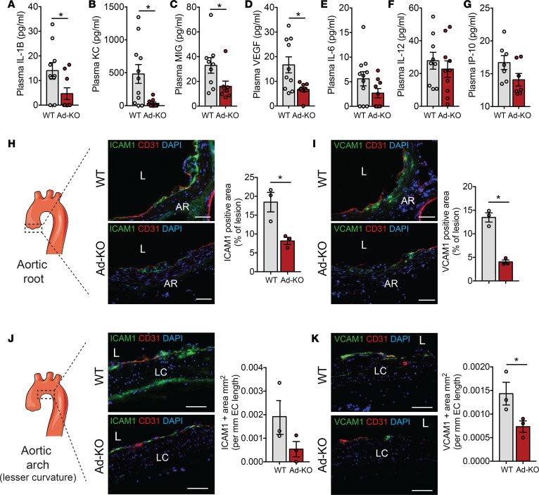
Figure 8. ANGPTL4 deficiency in adipose tissue (AT) reduces inflammation.
(A–G) Plasma concentration of indicated cytokines from WT mice and in mice with AT-specific knockout of ANGPTL4 (Ad-KO) mice fed a Western diet (WD) for 16 weeks measured by ELISA (n = 7–11). (H and I) Immunostaining showing the expression of ICAM-1 (H) and VCAM-1 (I) along with CD31 in section from aortic root atherosclerotic lesions of WT and Ad-KO mice fed a WD for 4 weeks (n = 3). The scheme on the left shows the region from which sections were taken. Panels on the right of each image show quantification of staining expressed as percentage of lesion area. AR, aortic root; L, lesion. (J and K) Immunostaining showing the expression of ICAM-1 (J) and VCAM-1 (K) along with CD31 in a section from the lesser (LC) curvature of aortic arch of WT and Ad-KO mice fed a WD for 4 weeks (n = 3). The scheme on the left shows the region from which sections were taken. Panels on the right of each image shows quantification of staining expressed as positive area per unit length of endothelium. Scale bars: 62 μm (H–K). All data are the mean ± SEM. *P ≤ 0.05 by comparison with data from Ad-KO with WT mice by unpaired t test.

