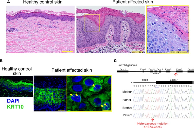
Figure 2. Histological and genetic features of the patient.
(A) Compared with healthy control skin (left panel), the patient’s affected epidermis, including the stratum corneum, was thicker (center panel). Parakeratosis, perinuclear vacuolization of keratinocytes, and marked reduction of keratohyalin granules were also noted (right panel). Scale bar: 100 μm. (B) WT KRT10 forms intermediate filaments in the cytoplasm of the suprabasal keratinocytes. In contrast, ectopic accumulation of KRT10 in the nucleus was observed in the affected epidermis (arrowheads). Scale bar: 50 μm (left and center panels), 10 μm (right panel). (C) A heterozygous splice-site mutation (c.1374-2A>G) in KRT10 was carried by the patient, whereas the unaffected family members were WT for this mutation.

