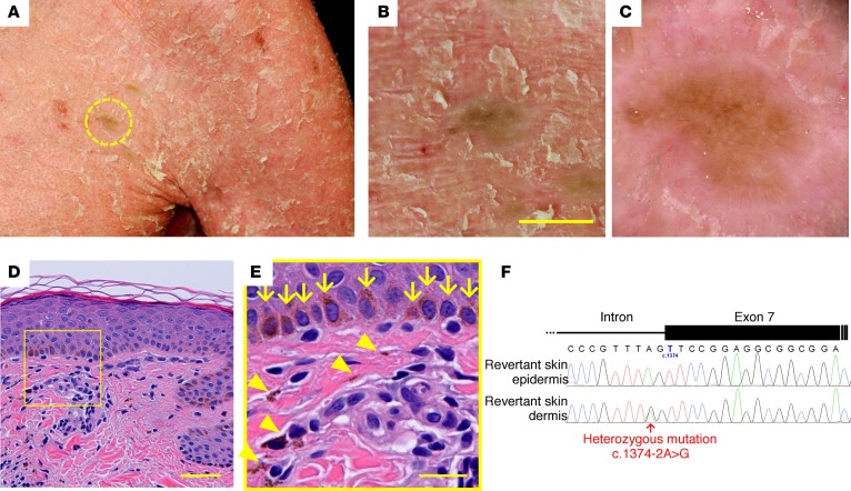
Figure 3. Identification of revertant spots.
(A–F) Gross (A and B), dermoscopic (C), histological (D and E), and genetic (F) features of the revertant spot on his left chest. (B) Note that no scales were observed on the revertant spot. Scale bar: 10 mm. (D) The revertant spot showed histological normalization. Scale bar: 50 μm. (E) High-power view of the square area in D. Basal melanosis (arrows) and incontinentia pigmenti histologica (arrowheads) were evident. Scale bar: 20 μm. (F) The splice-site mutation was lost in the revertant epidermis.

