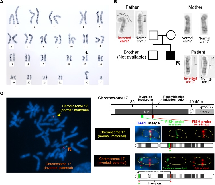
Figure 4. Detection of inv(17)(p13q12) and its breakpoints.
(A) G-banded karyotype analysis using peripheral blood lymphocytes from the proband revealed that the patient was heterozygous for a pericentric inversion (arrow), inv(17)(p13q12). (B) His unaffected father is also heterozygous with respect to inv(17)(p13q12), suggesting that the inversion itself is nonpathogenic. His mother is WT for the inversion. (C) Two-color FISH analysis using 2 BAC clone probes, RP11-141B22 (red) and RP11-259P8 (green), revealed a split red signal, suggesting that the breakpoint of the inversion is located in the RP11-141B22 locus.

