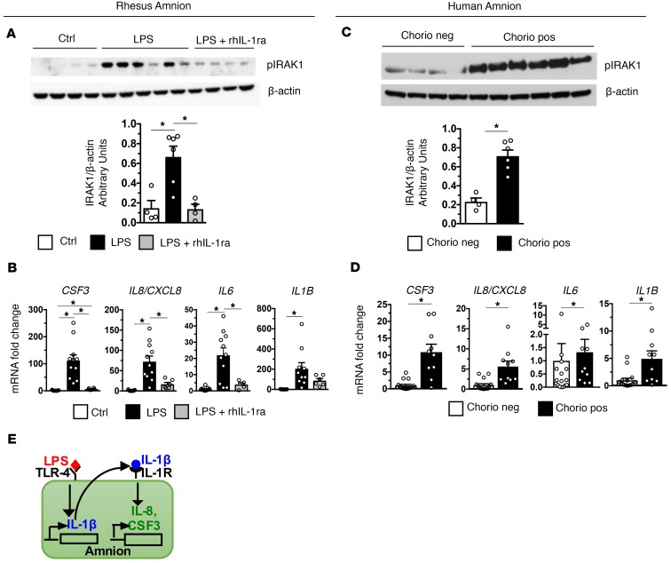
Figure 2. Activation of IRAK1 and induction of neutrophil chemoattractants is IL-1 dependent in the amnion.
Amnion was physically separated from chorion and decidua immediately after birth from rhesus and humans delivering preterm. (A) Representative immunoblots of rhesus amnion (n = 5) probed with anti–phospho-IRAK1 and β-actin and quantification of IRAK1 expression (Ctrl, n = 4; LPS, n = 6; LPS + rhIL-1ra, n = 4) are shown. (B) Expression of IA LPS–induced CSF3, IL8/CXCL8, and IL6 but not IL1β mRNA were inhibited by rhIL-1ra (Ctrl, n = 13; LPS, n = 11; LPS + rhIL-1ra, n = 6). (C) Representative immunoblots of human amnion (n = 5) probed with anti–phospho-IRAK1 and β-actin and quantification of IRAK1 expression (Chorio neg., n = 4; Chorio pos., n = 6) are shown. (D) CSF3, IL8, IL6, and IL1β mRNAs increased in human chorio cases (Chorio neg., n = 16; Chorio pos., n = 11). (E) Model demonstrating IL-1β downstream of TLR and CXCL8/CSF3 downstream of IL-1R in the amnion. Data are mean ± SEM, *P < 0.05 between comparators by Mann-Whitney test.

