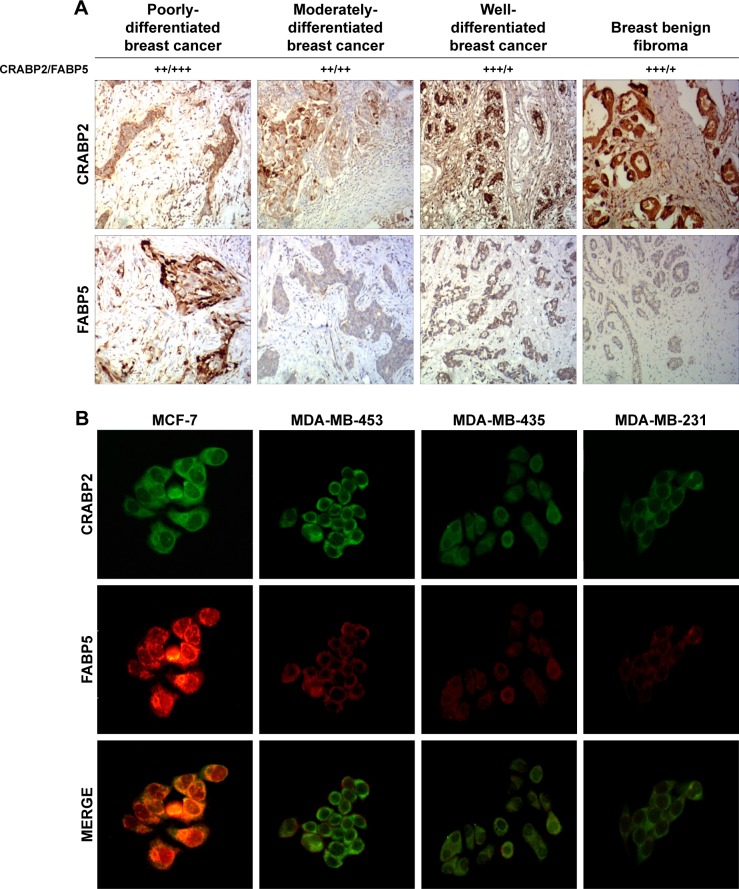
Figure 2.
Expression of CRABP2 and FABP5 in breast cancer tissues and cells.
Notes: (A) Expression of CRABP2 and FABP5 in breast cancer and breast benign fibroma was detected by immunohistochemistry. The staining patterns were scored as negative (−) if no immunolabeling was observed, weakly positive (+) if the labeling was faint, moderately positive (++) if the labeling was stronger, and strongly positive (+++) if the labeling was distinctly stronger than (++). Representative images are shown at 100× objective. (B) Expression of CRABP2 and FABP5 in different breast cancer cells was detected by immunofluorescence. Representative images are shown at 200× objective.

