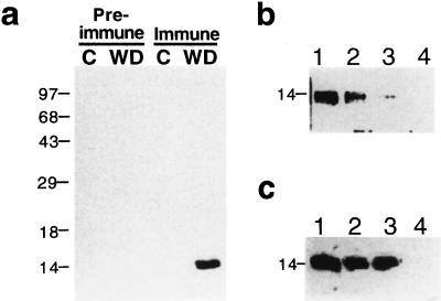
Figure 1.
Characterization of the anti-PvLEA-18 antibody. Bean seedlings grown in the dark for 5 d were transferred to well-irrigated or water-stressed vermiculite and harvested after 24 h of treatment. Total protein extracts were purified from roots, separated by SDS-PAGE, transferred to nitrocellulose, and incubated with antisera as follows: a, Immunodetection of PvLEA-18 in protein extracts obtained from control (C) or water-deficient (WD) tissues using immune, anti-PvLEA-18, or preimmune antisera. b, Competition of the antiserum by preincubation with different concentrations of purified PvLEA-18-GST fusion protein: 0 ng (lane 1), 50 ng (lane 2), 500 ng (lane 3), and 5 μg (lane 4). c, Competition analysis of the antiserum by addition of purified GST protein in different amounts: 0 ng (lane 1), 5 μg (lane 2), and 50 μg (lane 3). As a control, 5 μg of purified PvLEA-18-GST fusion protein was added in lane 4. Numbers at the left indicate the corresponding molecular masses in kD.

