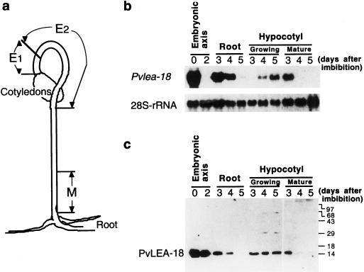
Figure 4.
Analysis of the accumulation of Pvlea-18 transcript and protein in roots and in different regions of the hypocotyl during seedling establishment. a, Schematic description of the hypocotyl regions used. E1 and E2, Hypocotyl-growing regions (E2 shows the highest elongation rate); M, nongrowing or mature zone. b, Northern-blot analysis of the Pvlea-18 transcript. Bean seeds were germinated in the dark and seedlings were harvested at different times (0, 2, 3, 4, and 5 d after imbibition). Five micrograms of total RNA was purified from different seed or seedling organs and from the hypocotyl regions indicated above, blotted on nylon membranes, and hybridized. Hybridization against a 28S-rRNA probe was used as an RNA-loading control. c, Western-blot analysis of the PvLEA-18 protein accumulation from total protein extracts obtained from the same samples as described in b. Numbers at the right indicate the corresponding molecular masses in kilodaltons. Proteins were separated by SDS-PAGE and transferred to nitrocellulose membranes before incubation with the immunopurified anti-PvLEA-18 antiserum.

