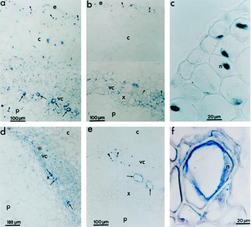
Figure 8.
Immunohistochemical detection of PvLEA-18 in protoxylem cells and nuclei from root and hypocotyl tissues using immunopurified anti-PvLEA-18 antibody. Tissues were dissected and soaked in paraffin as described in Figure 7. a and c, Sections from the hypocotyl-elongating region E1 obtained from water-stressed seedlings corresponding to ×2 and ×5 magnifications of Figure 7b, respectively. b, Section from the hypocotyl mature region obtained from water-stressed seedlings corresponding to a ×2 magnification of Figure 7h. d, Section from the emerging hypocotyl obtained from well-irrigated seedlings after 36 h of imbibition. e, Section from a root obtained from well-irrigated seedlings grown as described in Figure 7. f, Detail of a protoxylem cell from the mature region of the hypocotyl of a 6-d-old well-irrigated seedling. Arrows indicate protoxylem cells and arrowheads indicate nuclei. c, Cortex; e, epidermis; p, pith; vc, vascular cylinder; x, xylem.

