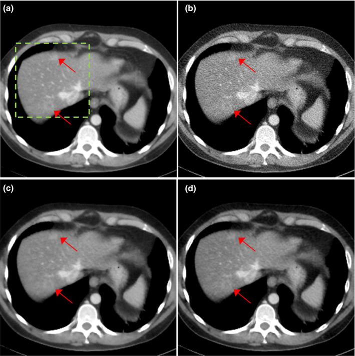
Figure 7.

Patient of L291 with metastatic lesions. The reconstructed images using (a) regular dose FBP, (b) quarter dose FBP, (c) quarter dose TV and (d) quarter dose proposed method were compared. Intensity range is 40 ± 200 HU. Metastatic lesions are pointed by red arrows.
