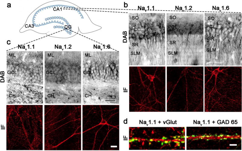
Figure 2.

Nav subtypes are expressed in soma, dendrites and axons of hippocampal CA1 and DG. A. Schematic diagram of CA1, CA3, and dentate gyrus (DG) in rat hippocampus. B-C. High magnification light microscopic photomicrographs (top) of Nav 1.1 (left), Nav 1.2 (middle), and Nav 1.6 (right) in rat CA1 (B) and DG (C). Nav labeling is evident in stratum oriens (SO), pyramidal cell layer (PCL), stratum radiatum (SR), and stratum lacunosum-moleculare (SLM) in CA1 (B, top) and in molecular layer (ML), granular cell layer (GCL), and central hilus (CH) in DG (C, top). Bar 200 μm. Examples of immunofluorescence localization of Nav 1.1 (left), Nav 1.2 (middle), and Nav 1.6 (right) in rat hippocampal cultures from CA1 (B, bottom) and DG (C, bottom). Bar 15 μm. D. Immunofluorescence images showing double labeling of Nav 1.1 (magenta) with excitatory axon terminal marker vesicular glutamate transporter (vGlut; green, left) and inhibitory axon terminal marker glutamic acid decarboxylase 65 (GAD 65; green, right). Bar 5 μm. See supplemental figure 2 for red-green images.
