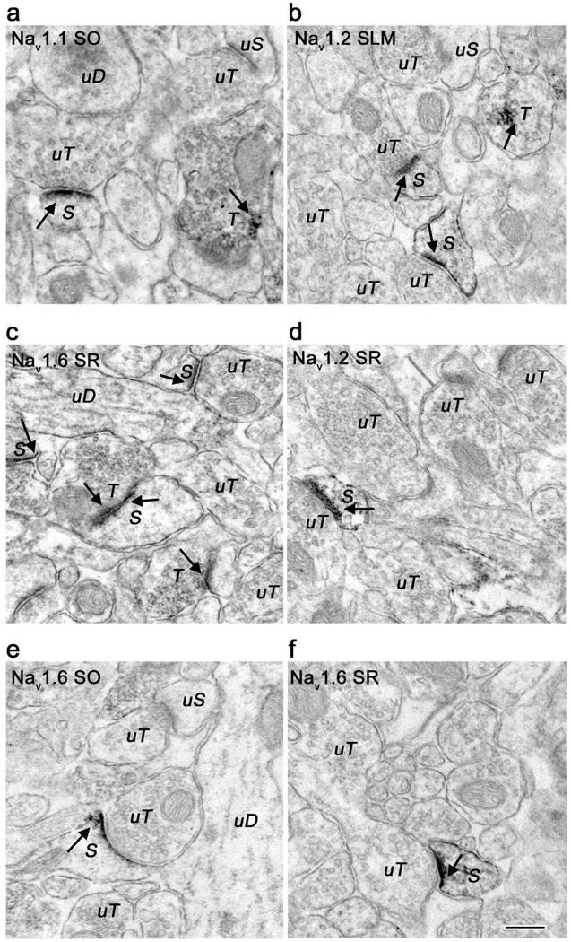
Figure 4.

Electron micrographs showing localization of Nav 1.1, Nav 1.2, and Nav 1.6 immunoreactivity in dendritic spines. A. Nav 1.1 immunoreactive dendritic spine (S) forms a synapse with an unlabeled terminal (uT) in CA1 stratum oriens (SO). Discrete labeling was seen predominantly in the spine head. Nearby unlabeled dendrite (uD) and terminal (uT) as well as a labeled terminal (T) are shown for comparison. B, D. Nav 1.2 immunoreactivity in CA1 stratum-lacunosum-moleculare (SLM) and stratum radiatum (SR) in labeled spine heads and spine necks (S) forming synapses with unlabeled terminals (uT). Two labeled branching spines are seen in panel B. Nearby labeled (T) and unlabeled terminals (uT) are shown. C, E, F. Nav 1.6 immunoreactivity in dendritic spines in CA1 SO, SR, and SLM. Multiple labeled spines (S) are seen in panel C forming synapses with labeled (T) and unlabeled (uT) terminals. Immunoreactivity shown with arrows. Bar 250 nm.
