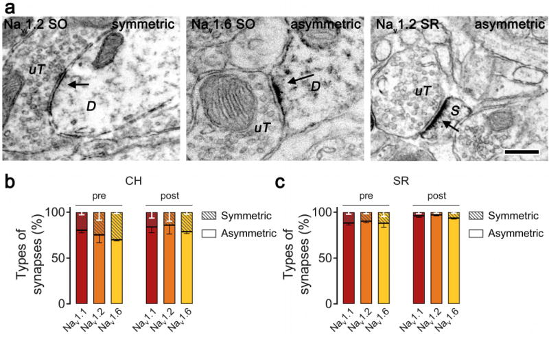
Figure 7.

Nav immunoreactivity is predominantly associated with asymmetric synapses in the stratum radiatum (SR) and central hilus (CH) subregions. A. A Nav 1.2 unlabeled terminal (uT) is seen making a symmetric synapse with a labeled dendrite (D; left) and a labeled spine (S; right). A Nav 1.6 unlabeled terminal is seen making an asymmetric synapse with a labeled dendrite (D; middle). Bar 250 nm. B, C. Quantitative analysis showing pre- and post-synaptic structures containing Nav immunoreactivity is largely associated with asymmetric synapses in the SR (B) and CH (C). Data were analyzed from 15 micrographs obtained from three rats per experimental group; n = 265 to 316 synapses in CH and 424 to 540 synapses in SR.
