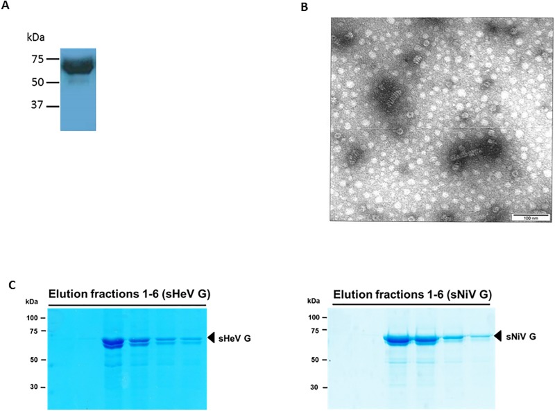
Fig 1. Purification and detection of ELISA antigens.
A. Immunoblot of purified his-tagged NiV N expressed in Escherichia coli. The identity of the NiV N protein was confirmed by anti-N monoclonal antibody F45G4 using enhanced chemiluminescence. B. Negative contrast electron microscopy of an inclusion body fraction of semi-purified NiV N antigen. The specimen grids were examined in a Philips CM 120 transmission electron microscope, operating at an accelerating voltage of 80 kV. Micrographs were taken between 28,000X–45,000X using Kodak Electron Microscope Film 4489. The negatives were scanned using an Epson Perfection 3200 photo scanner and enlarged 2.5X. C. Coomassie staining of purified recombinant sHeV G and sNiV G protein elution fractions. Purification of Strep-tagged sHeV G (A) and sNiV G (B) from Leishmania tarentolae cell lysates was performed via Strep-tag affinity chromatography. Elution fractions were separated by 10% SDS-PAGE under reducing conditions and visualized by Coomassie staining.

