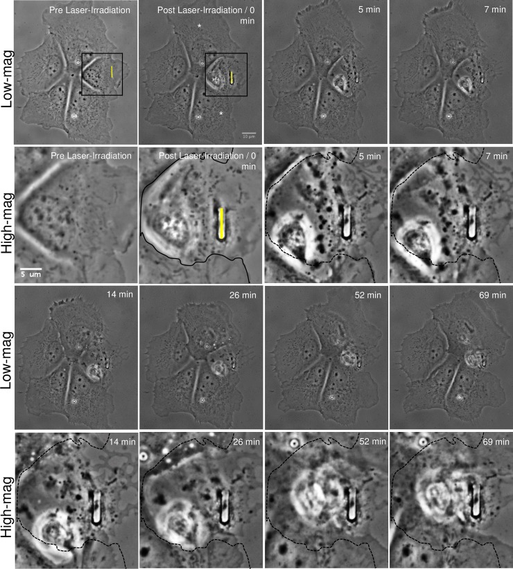
Fig 3. Phagocytosis of a laser-irradiated astrocyte within a cell cluster.
An astrocyte within a five-cell cluster is lysed via laser nanoablation along the yellow ROI, visible in the first two images. Contrast enhanced, higher magnification images of the irradiated region are displayed in the second and fourth rows labeled “High-mag”. High magnification images correspond to the region within the outlined box in both pre and post laser-irradiated cell images. Immediately following laser exposure, the targeted cell shows visible signs of death including membrane retraction from the dish surface, phase paling of the nucleus, and phase dark material aggregating around the laser-irradiated site. Two cells closest to the targeted cell (white asterisks) share significant membrane contact with the irradiated cell. Between 5–69 minutes, the two astrocytes respond by extending their membranes toward the dead cell debris that they eventually surround and engulf. To better define the advancement of the responding astrocytes’ lamellae into the region of the irradiated cell, the boundary edge of the irradiated cell is traced with a solid black line in the post laser-irradiated region. This line is represented as a dashed black line in the subsequent high-magnification images. As the lamellae advances, vesicles form within the two responding cells starting 14 minutes following laser irradiation. The two other cells within the cluster, not in close proximity (and not sharing extensive membrane contact with the irradiated cell), do not exhibit any visible response following laser irradiation of the targeted cell.

