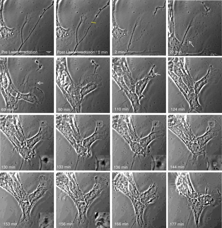
Fig 5. Astrocyte phagocytosis of dissected neurite debris.
Phase contrast images (contrast enhanced) depicts the uptake of a neuron process by an astrocyte residing to the left of the process. Laser exposure to an axon (yellow ROI) results in thinning, blebbing, and full dissection of the growth cone. Following the dissection, the detached growth cone continues to actively move. At 22 minutes post laser irradiation, the neighboring astrocyte physically connects to the damaged process and begins to pull on debris (white arrow). The responding astrocyte (asterisk) continues to move along the damaged neurite between 57 and 69 minutes post laser irradiation in the direction of the growth cone. The astrocyte reaches the active growth cone 90 minutes following laser irradiation. Magnified insets highlight the intake of the growth cone, including phase dark material at the periphery of the cell membrane, engulfment of the debris, formation of endocytic vesicle, and movement of the debris toward the cell center. 124 min following laser irradiation, the leading edge is reformed followed by cell migration in the opposing direction, revealing no debris from growth cone remaining in the previously occupied region.

