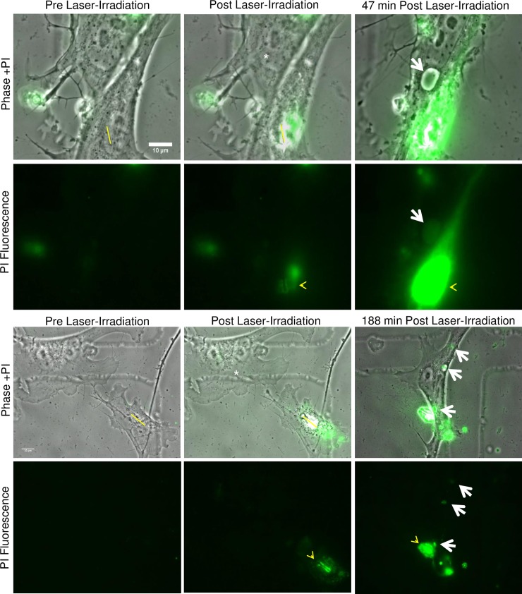
Fig 7. Propidium iodide stained nuclear material is incorporated into endocytic vesicles of responding astrocytes.
Prior to laser exposure, PI was added to the medium surrounding primary astrocytes. Minimal intracellular PI fluorescence was observed within living cells, as visualized in the “Pre Laser-Irradiation, PI Fluorescence” images. After laser irradiation, PI is incorporated into the exposed DNA of the targeted cell along the yellow ROI, and was visualized as an increase in fluorescence intensity in “Post Laser-Irradiation” images (yellow arrow heads). Fluorescence of the exposed nuclear material continues to increase, producing a bright signal in the region of the targeted nucleus. This was visible 47 minutes post fluorescence in the first example and 188 minutes in the second example. Large endocytic vesicles were observed within the responding astrocytes (white asterisk) 47 and 188 minutes following laser-irradiation. Overlay images of phase and fluorescence confirm that the PI-stained nuclear material was located within the large endocytic vesicles (white arrows).

