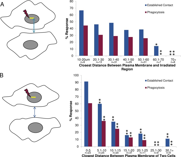
Fig 9. Distance dependent response of isolated astrocytes.
A. Response of cells based on the closest distance between the plasma membrane of the responding astrocyte and the laser-irradiated region. A. Schematic diagram of distance measured between two observed cells. The blue line corresponds to the shortest distance between location of the laser ROI (yellow line) and the plasma membrane of the responding astrocyte. This is equivalent to the shortest distance material released from the irradiated region must diffuse to be sensed by the responding astrocyte. A higher rate of establishing cell-to-cell contact and subsequent phagocytosis was observed for cells situated closer to each other. A significant decrease in the responding astrocyte’s ability to establish contact with an irradiated cell occurred for cells separated by 60–70 μm (14%, p<0.05), and 70+ μm away (0%, p<0.01), as compared to 67% established contact response observed when the irradiated/lysed cell and the responding cell are 10–20 μm apart. Similarly, a significant decrease in phagocytic response was observed when the cells were 60–70 μm and 70+ μm microns apart (p<0.01) with a 0% phagocytic response, as compared to 44% observed for the 10–20 μm distance. B. Response based on the closest distance between plasma membranes of the laser-irradiated cell and the responding cell. C. Schematic diagram of the closest distance measured between the plasma membrane of two cells (blue line). As in Fig 9B, a higher rate of establishing cell-to-cell contact and phagocytosis was observed for cells closer to each other. A significant decrease (p<0.01) in the responding astrocyte’s ability to establish contact occurred for distances greater than 5 μm, when compared to those distance in the 0–5 μm range (91%).

