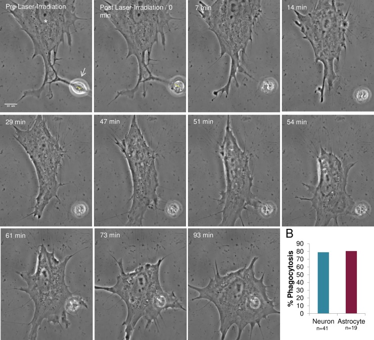
Fig 12. Phagocytosis of laser-irradiated neurons by neighboring astrocytes.
A. Neurons from mixed primary cortical cultures that shared membrane contact with neighboring astrocytes were targeted in the nucleus (yellow ROI). Neurons damaged by laser exposure rounded and released significant cell debris as visualized in the Post Laser-Irradiation image. The responding astrocyte (white asterisk) engulfed the cellular debris 56–75 minutes following irradiation. This response is similar to phagocytosis of laser-irradiated astrocytes. B. Astrocyte responses to laser-killed neurons are similar to responses to laser-killed astrocytes. Results are similar to astrocytes sharing a membrane connection, with rates of phagocytosis of lysed astrocytes at 81% (15/19), and lysed neurons at 79% (32/41).

