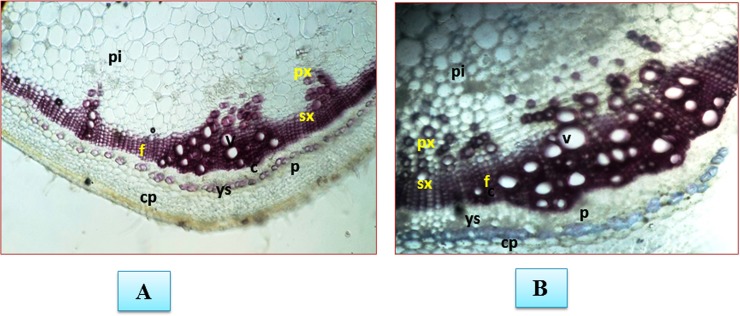
Fig 7. Assessment of plant defense response in the form of lignification.
The pink colour shows the amount of lignified tissues. The shoot tissues between the second and third nodes from Fol challenged plants were collected after 3 weeks post inoculation. The photograph were taken after staining with phloroglucinol-HCl A. Control B. Fol challenged plants. Transverse section of tomato stem stained with pholoroglucinol-HCl at 2nd internode showing the lignified tissues in pink colour px = primary xylem; sx = secondary xylem; f = xylem; pi = pith; p = phloem; c = cambium; v = vessel; cp, cortical parenchyma. In control sample the amount of lignin deposition is less. The Fol challenged stem showed the intense pink coloration of the lignified tissues and the high intensity of pink colour in samples represent the high amount of lignified material deposited.

