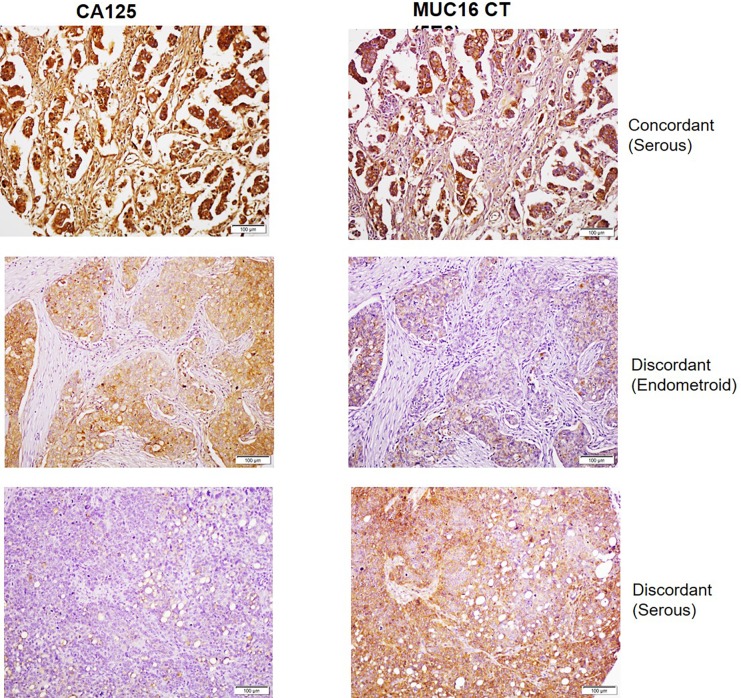
Fig 5. Comparative analysis of CA125 and MUC16 CT staining in human ovarian cancer cases by IHC.
Duplicate ovarian cancer tissue microarrays (OV1004, BIOMAX) were processed for IHC staining as described in the Materials and Methods section and with primary antibodies CA125 (M11) and MUC16 CT (5E6). Representative cases of concordant and discordant staining are indicated. The top and bottom panels represent serous subtypes which exhibited intense staining with both CA125 and MUC16 CT mAbs. The middle panel represents the endometroid subtype that exhibited positive reactivity with CA125 but demonstrated comparatively lower staining with MUC16 CT mAb.

