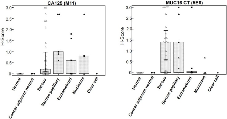
Fig 6. Box plot comparing CA125 and MUC16 CT staining across various histologic types of ovarian cancer.
MUC16 immunoreactivity was higher in the serous and serous papillary adenocarcinoma tissues as compared to other types. The CA125 mAb also exhibited reactivity with endometroid and mucinous types while CT mAb failed to detect these types efficiently. No staining is observed in normal or cancer adjacent normal ovarian tissue with either CA125 or MUC16 CT mAbs.

