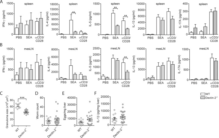
Fig 7. Dectin-2 signaling is required for induction of a Th2 response during S. mansoni infection.
WT and Dectin-2−/− mice were infected with S. mansoni. After 8 wk of infection, cells from spleens (A) or mLNs (B) were restimulated with SEA or anti-CD3/CD28 for 72 h, and cytokine levels were analyzed in supernatants by Luminex or ELISA. Bars represent mean ± SEM of combined data of at least 2 or 3 independent experiments with 5 to 10 mice per group. (C) Granuloma sizes around eggs trapped in the liver of 8-wk–infected mice were assessed in Masson blue–stained liver sections. Data are based on 10 mice per group. Number of worms (D) and liver and intestinal eggs (E) in mice infected with S. mansoni for 8 wk. (F) IL-1β protein levels in livers of mice infected with S. mansoni for 8 wk. **P < 0.01 and ***P < 0.001 for significant differences relative to the control mice based on unpaired analysis (unpaired Student t test). Underlying data can be found in S1 Data. CD3, cluster of differentiation 3; IL-1β, interleukin 1β; mLN, mesenteric lymph node; SEA, soluble egg antigen; Th2, T helper 2; WT, wild-type.

