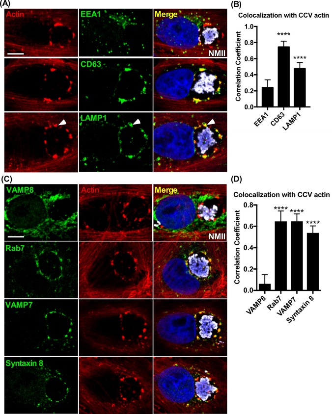
Fig 1. Actin patches on the CCV membrane are enriched for late endocytic vesicles and fusion regulatory proteins.
(A and B) Colocalization of endosomal components with CCV actin patches. Vero cells fixed at 3 dpi and stained for F-actin, early endosomes (EEA1+), late endosomes (CD63+), or lysosomes (LAMP1+). CCV actin patches cluster with late endocytic markers. Clustering with large, individual vesicles is also seen (white arrows). (C and D) Colocalization of fusion regulatory proteins with actin patches. Histograms depict the means ± SD of ≥ 60 cells for at least 3 independent experiments. Statistical significance was determined using Student’s t-test (****P <0.0001). Colocalization was determined using Pearson’s correlation coefficient. NMII, C. burnetii Nine Mile phase II strain. Scale bar, 5 μm.

