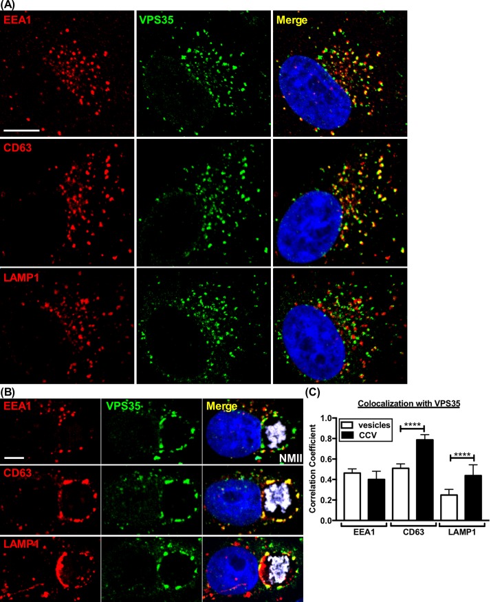
Fig 5. Retromer colocalizes with CD63+ vesicles that cluster with CCV actin patches.
(A and C) Vero cells were immunostained for retromer (VPS35+), early endosomes (EEA1+), or late endosomes/lysosomes (CD63+, LAMP1+). EEA1+ and CD63+ vesicles colocalize similarly with VPS35, while LAMP1+ vesicles are much less colocalized. (B and C) Vero cells 3 dpi, stained as in (A). Clustered CD63+ vesicles on the CCV membrane have high colocalization with VPS35 compared to EEA1+ vesicles. LAMP1+ vesicles on the CCV membrane also show increased colocalization with VPS35. Colocalization analysis of vesicles and CCVs was determined using Pearson’s correlation coefficient. Graphs represent the means ± SD of ≥ 60 cells from at least 3 independent experiments. Statistical significance determined by Student’s t-test (****P <0.0001). NMII, C. burnetii Nine Mile phase II strain. Scale bar, 5 μm.

