
Figure 2.
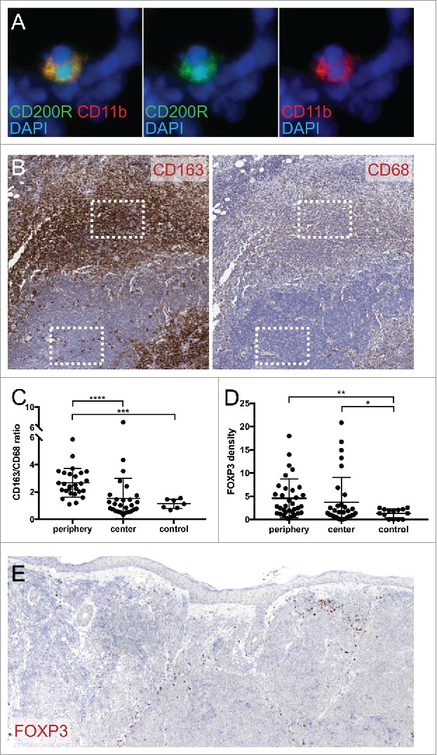
MCC is associated with immunosuppressive immune infiltrate. (A) CD200R and myeloid marker CD11b immunostaining in fresh-frozen MCC tumor. Original magnification 400x. (B) M2 marker CD163 and macrophage marker CD68 staining in MCC tumor. Representative sampling areas for tumor periphery and tumor center used for staining quantifications (boxes). Original magnification 50x. (C) Mean CD163/CD68 staining density ratio for MCC tumor periphery, tumor center, and normal skin control. ****, P<0.0001 by paired t-test; ***, P<0.0001 by Welch's unpaired t-test. (D) Mean Treg marker FOXP3 staining density for MCC tumor periphery, tumor center, and normal skin control. **, P<0.0005; *,P<0.05. (E) FOXP3 staining in MCC tumor. Original magnification 100x.