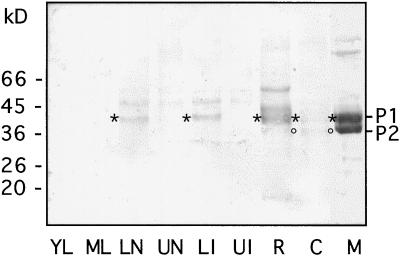
Figure 6.
Detection of P-type isoPOXs in extracts of different plant organs and BY-2 cells and in cell-suspension culture medium. Soluble proteins were extracted from young leaves (lane YL), mature leaves (lane ML), lower stem nodes (lane LN), upper stem nodes (lane UN), lower stem internodes (lane LI), upper stem internodes (lane UI), roots (lane R), and 7-d BY-2 cells (lane C). Ten micrograms of these extracts was analyzed by western blotting together with an aliquot of cell-suspension culture medium (lane M) containing 1.2 μg of protein. Polyclonal antibodies raised against P-type isoPOXs were used; the asterisks and circles indicate the positions of signals corresponding to P1 and P2, respectively. The control with preimmune serum did not show any signal.

