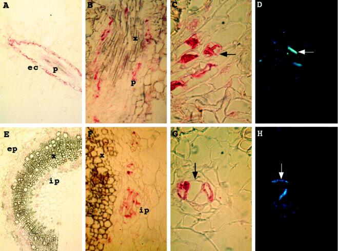
Figure 7.
Immunocytolocalization assays using anti-P-type POX antibodies. A, Root longitudinal section; B, close-up of root longitudinal section; C, close-up of transversal root section; D, same as C but stained with aniline blue; E, stem transversal section; F, close-up of stem section shown in E; G, close-up of stem transversal section; and H, same as G but stained with aniline blue. ec, Epidermal cells; ep, external phloem; ip, internal phloem; p, phloem; and x, xylem. Arrows indicate the localization of walls visualized by fluorescence with aniline blue.

