Figure 2.
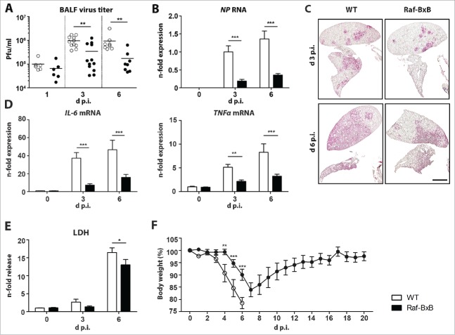
Comparative analysis of IAV replication in tumor-bearing and wildtype mice. C57Bl/6 WT and Raf-BxB mice were infected with 500 particles of IAV PR8 and at indicated days post infection, BALF and lung tissue were studied. (A) Virus titers presented as pfu/ml were determined in BAL samples by standard plaque assay. (B) RNA levels of viral NP were determined by qRT-PCR. The mean value of infected WT mice at day 3 p.i. was arbitrarily set to one. (C) Viral spread within the lungs of infected WT or Raf-BxB tumor-bearing mice was analyzed at day 3 and 6 post infection by immunohistochemistry staining of IAV NP protein. Bars = 2000 μm. (D) mRNA levels of pro-inflammatory cytokines IL-6 and TNFα were determined by TaqMan qRT-PCR. Values of cytokines in uninfected WT mice were set as unity. (E) Relative LDH content in BALF of WT and Raf-BxB mice. The mean value of non-infected WT mice was set to one. (F) Bodyweight loss of infected WT and tumor-bearing Raf-BxB mice after IAV infection. Mean values ± SEM of two experiments with ≥5 animals per group in each experiment are presented.

