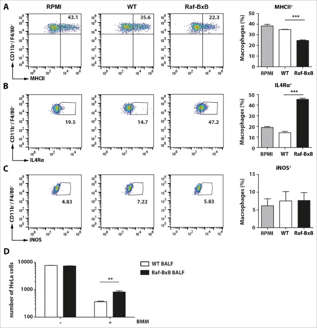
Figure 4.
Incubation of bone marrow-derived macrophages with BALF from WT or tumor-bearing Raf-BxB mice. Bone marrow-cells from WT mice were isolated and differentiated to bone marrow-derived macrophages (BMM) (CD11b+/ F4/80+) by incubation with M-CSF for 5 days. (A, B, C) Then, the cells were re-cultured for 48h in the presence of BALF from uninfected WT or Raf-BxB mice or RPMI medium as control and analyzed for surface expression of MHCII (A) or IL4Rα (B) or intracellular expression of iNOS (C) by flow cytometry. Representative dot-blot images underlining the gating strategy are exemplarily shown. (D) BMM were pre-activated over night with 1 µg/ml LPS and afterwards co-cultured with mitomycin-treated HeLa cells in presence of BALF derived from WT or Raf-BxB mice (E/T = 20/1). 48h post co-culture, the absolute number of remaining HeLa cells was determined via flow cytometry by usage of cell counting beads. Mean values ±SEM are shown. BALF was isolated and pooled from at least 3 different mice . Bone marrow cells were pooled from 3 animals.

