
Figure 4.
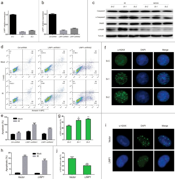
Knockdown of LINP1 increased IR-induced cell apoptosis and delayed repair of DNA double-strand breaks in cervical cancer cells treated with radiation a and b. QRT-PCR assays showing LINP1 silencing efficiency using siRNA or shRNA. c. Expression of caspase3, cleaved caspase3, PARP and cleaved PARP detected by western blotting in Hela S3 cells expressing LINP1 or control siRNAs 24 hours after treated with or without 6 Gy irradiation. d. Apoptosis was detected by flow cytometry in Hela S3 cells expressing control or LINP1-specific shRNAs 24 hours after treated with 6 Gy IR. e. The apoptotic rate was calculated as the percentage of Annexin V-APC-positive cells. f. Immunofluorescence stain visualizing IR-induced γ-H2AX foci in LINP1 silencing and control Hela S3 cells 24 hours after treatment. g. Quantification of the number of γ-H2AX foci positive cells expressing LINP1 or control siRNAs 24 hours after irradiation. h. The apoptotic rate of SiHa cells expressing control vector or LINP1 24 hours after treated with or without 6 Gy irradiation. i. Immunofluorescence stain detecting α-H2AX foci in LINP1-overexpressing and control SiHa cells 24 hours after 6 Gy IR treatment. j. Quantification of the number of γ-H2AX foci positive cells expressing control vector or LINP1 24 hours after exposure to irradiation. Cells with 5 or more γ-H2AX foci were counted as unrepaired cells or γ-H2AX foci positive cells. Si-C, cells expressing Ctrl-siRNA; Si-1, cells expressing LINP1-siRNA1; Si-2, cells expressing LINP1-siRNA2. PcDNA3.1 (+) plasmid transfected SiHa cell lines were used as controls; LINP1 indicates LINP1-overexpressing SiHa cells. Error bars, s.d. *P < 0.05 by two-tailed Student's t test; n = 3 independent cell cultures.