Figure 5.

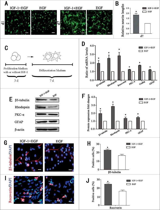
The differentiation potential of RPCs toward retinal neurons was enhanced by IGF-1 pretreatment. (A-B) Fluorescent micrographs of the GFP+ RPCs cultured in differentiation culture for the indicated times. The results showed that the cells in the IGF-1 plus EGF-pretreated RPC cultures exhibited longer neurites than in the EGF group. (C)Schematic diagram of the detection of the influence of IGF-1 on the differentiation of RPCs. RPCs were cultured in proliferation medium with IGF-1+EGF or EGF for 3 days, and then the cells were cultured in differentiation medium for 7 days. (D) The qPCR results showed that the expression levels of β3-tubulin, rhodopsin, recoverin, PKC-α, Cralbp and GFAP in IGF-1+EGF group were obviously increased compared to the EGF group. (E) Western blot analysis showed that the expression levels of β3-tubulin, rhodopsin, PKC-α, and GFAP were enhanced when RPCs pretreated with IGF-1+EGF compared with EGF. (F) Western blot protein expression signal revealed that the IGF-1+EGF pretreated RPCs expressed more β3-tubulin, rhodopsin, PKC-α, and GFAP protein than the EGF group. (G-J) The percentages of β3-tubulin-positive cells and recoverin-positive cells were significantly increased in the IGF-1+EGF group compared to the EGF group. *P ≤ 0.05 (Student's t-test). Scale bars: A: 100 μm, F: 25 μm.
