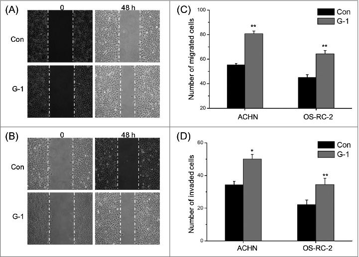
Figure 2.

Activation of GPER promoted the in vitro migration and invasion of RCC cells. (A) Confluent monolayers of ACHN cells were scraped by a pipette tip to generate wounds and then were cultured. Representative images of wounds at 0 and 48 h in the absence or presence of 1 μM G-1; (B) Representative images of wounds of OS-RC-2 cells at 0 and 48 h in the absence or presence of 1 μM G-1; ACHN or OS-RC-2 cells were allowed to migrate or invade transwell chambers into the under-side of the filter for the indicated times in the presence or absence of 1 μM G-1. The migrated cells were fixed, stained, and photographed. The migrated cells were fixed, stained, and photographed. The number of migrated (C) or invaded (D) cells were compared with the control. Data are presented as means ± SD of three independent experiments. *P < 0.05 compared with control; **P < 0.01 compared with control.
