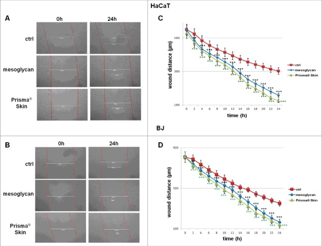
Figure 3.

Evaluation of migration rate of human keratinocytes and fibroblasts in presence of mesoglycan and Prisma® Skin. Representative images of Wound Healing assay on HaCaT (A) and BJ (B) cells treated or not with sodium mesoglycan and Prisma® Skin 0.3 mg/ml. Results of Wound Healing assay analysis for HaCat (C) and BJ (D) cells. The migration rate was determined by measuring the wound closure by individual cells from the initial time to the selected time-points (bar of distance tool, Leica ASF software). The data represent a mean of 3 independent experiments ± SEM, their statistical significance were evaluated using Student's t-test, assuming a 2-tailed distribution and unequal variance. **p < 0.01; ***p < 0.001.
