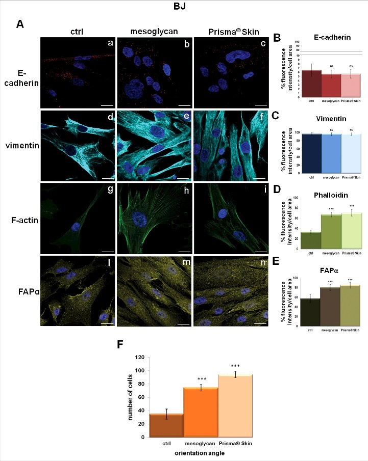
Figure 6.

Characterization of cytoskeletal organization and activation of fibroblasts. (A) Immunofluorescence analysis to detect E-cadherin (panels a, b, c), vimentin (panels d, e, f), F-actin (panels g, h, i), FAPα (panels l, m, n) treated with mesoglycan and Prisma® Skin for 48 h. Quantification of (B) E-cadherin (a, b, c), (C) vimentin (d, e, f), (D) phalloidin (g, h, i) and (E) FAPα (l, m, n) as percent of fluorescence intensity per cell area. (F) Cell distribution was determined by calculating the percentage of cells arranged in parallel for each acquired region ( ± 100 of the mode angle). Nuclei were stained with DAPI. Magnification 63 × 1.4 NA. Bar = 10 µm. All the results are representative ± SEM of 3 experiments based on Student's t-test, assuming a 2-tailed distribution and unequal variance. ***p < 0.001, ns p > 0.05.
