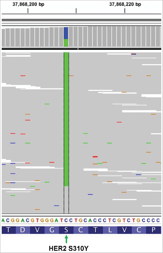
Figure 3.

The Intergrative Genomics Viewer (IGV) screenshots displayed the reads from ctDNA sequencing and revealed the harboring of HER2 S310Y. Different nucleobase types were presented with different colors. Adenine (A) is presented by green, cytosine (C) is indicated by blue, guanine (G) is yellow, and thymine (T) is red. The green column in the middle indicates position of the mutated nucleobase (929C>A). The mutation of HER2 S310Y in terms of nucleobase and amino acid level occurred as 929C>A and Serine310 to Tyrosine, respectively [NM_004448.3(HER2):c.929C>A(p.Ser310Tyr)].
