Figure 8.

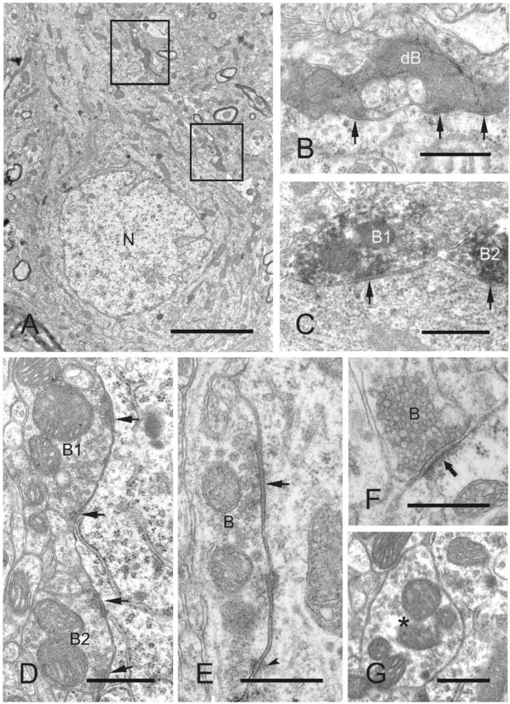
Electron microscopic analysis of symmetric (inhibitory) and asymmetric axosomatic synapses on layer V pyramidal neurons in naïve sensory-motor cortex (control, n = 2 rats) and undercut and contralateral cortex (undercut, undercut contra, n = 3 rats). 25 micrographs examined from each of 2 specimens/rat. A: Electron micrograph of a layer V pyramidal neuronal soma from undercut neocortex showing the nucleus (N), cytoplasm, and degenerated axonal boutons (boxed areas, one of which is shown enlarged in B) making synapses with the soma. B: Higher magnification of a degenerated axonal bouton (enpassant) exhibiting multiple synaptic contacts (arrows) with the soma. C: Electron micrograph of GAD65 immuno-positive axonal boutons (dark reaction product; B1, B2) exhibiting symmetric synapses (arrows) with the soma of a layer V pyramidal neuron. D: Electron micrograph of axonal boutons (B1, B2) making multiple symmetric synapses (arrows) with the soma of a neocortical layer V pyramidal neuron from a control rat. E: Electron micrograph of an axonal bouton (B) exhibiting a symmetric synapse (arrow) and puncta adherentia (arrowhead) with the soma of a layer V pyramidal neuron within the undercut neocortex 3 weeks after UC. F: Electron micrograph of an asymmetric synapse (arrow) between an axonal bouton (B) and the soma of a layer V pyramidal neuron from a control animal. G: Electron micrograph of a bouton (asterisk) containing mitochondria and synaptic vesicles adjacent to a layer V pyramidal neuronal soma, but without a synapse at this section plane. Scale bars: A, 4 μm; B–F, 500 nm; G, 1 μm.
