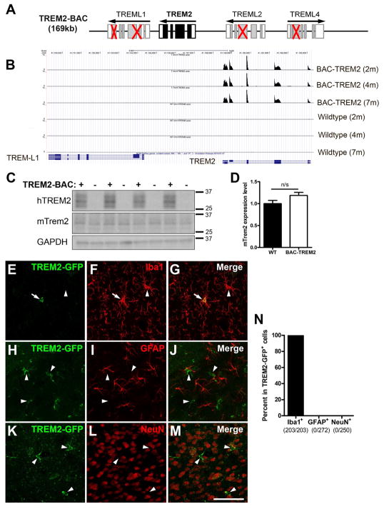
Figure 1. Generation and characterization of BAC-TREM2 mice.
(A) Schematic representation of the modification of TREM2-BAC. Red crosses indicate the deleted exons in TREM-like genes in the BAC construct.
(B) UCSC genome browser track showing read coverage at the human TREM2 locus in TREM2 transgenic and wildtype animals.
(C) Western blot was performed using the hippocampal lysates from 1.5 month-old WT and BAC-TREM2 mice with human TREM2 and mouse Trem2-specific antibodies. GAPDH served as a loading control.
(D) The band intensity of Western blots was quantified using Image J and shown as ratio of mTrem2/GAPD (n = 4).
(E–N) Brain sections from 1.5–2 month-old BAC-TREM2-GFP mice were double stained with GFP and cell-specific markers for microglia (Iba+, E–G), astrocytes (GFAP+, H–J), or neurons (NeuN+, K–M). Representative cortical images showed that BAC-TREM2-GFP colocalized with Iba1 (E–G) but not with GFAP (H–J) not NeuN (K–M). Bar = 100μm. (N) GFP+ cells were examined for the colocalization with cell-specific markers and presented as percent double labeled cells over GFP+ cells. The numbers below X-axis indicate the number of cell counted.

