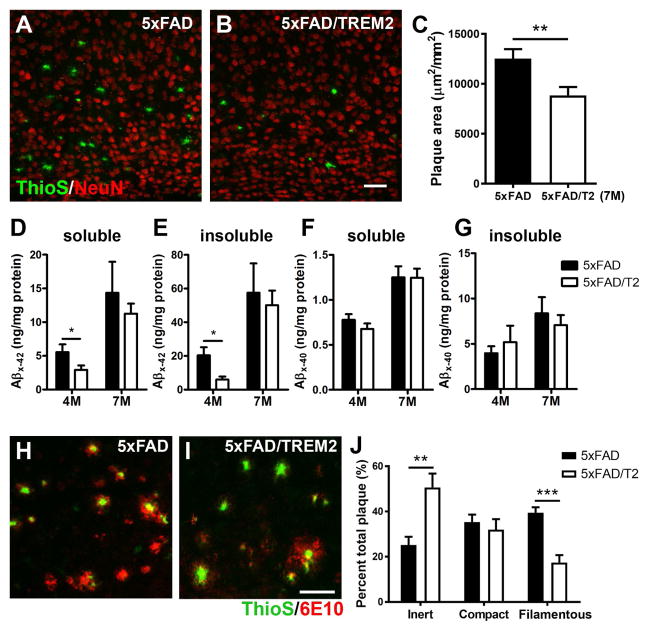
Figure 2. Increased TREM2 gene dosage ameliorates amyloid pathology and remodels amyloid plaque types.
(A–C) Matched brain sections from 7-months-old 5xFAD (A) and 5xFAD/TREM2 mice (B) were stained with ThioS (green) and NeuN (red) to visualize the amyloid plaques in the cortex. Z-stack confocal images (20X) were utilized to measure total plaque area in the field using ImageJ. The results are presented as ThioS+ plaque area (μm2) per mm2 of the cortical area (C). n = 7 per genotype, **p < 0.01. Bar = 50μm.
(D–G) The levels of soluble and insoluble Aβ42 (D and E) and Aβ40 (F and G) in the cortex of 4 and 7-month-old mice were measured by ELISA. n = 6 per genotype, *p < 0.05.
(H–J) Matched brain sections from 7-month-old 5xFAD (H) and 5xFAD/TREM2 mice (I) were stained with ThioS and anti-Aβ antibody (6E10). Z-stack confocal images (40X) were utilized to quantify 3 different forms of plaques using ImageJ (J). A total of 502 plaques were analyzed and are presented as mean ± SEM. n = 4 per genotype, **p < 0.01, ***p < 0.001. Bar = 50μm.

