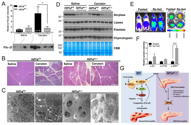
Fig. 2. HIF1α regulates pancreatic physiologic and pathophysiologic functions.
(A) Fib-γD immunoblot (and relative intensity) of the insoluble protein fraction isolated from pancreata after saline or cerulein administration. (B) Histology (bar=200μm), and (C) Transmission electron microscopy (bar=5μm) images of pancreata from Hif1aF/F or Hif1aAc−/− mice. (D) Total pancreas lysates were blotted with antibodies to the indicated antigens. (E) Bioluminescence of ODD-luc mice and pancreata after 14h-fasting or re-feeding. (F) Relative protein levels of immunoblots of pancreata lysates (see Fig. 11S for blots). (G) Summary of findings. HIF1α contributes to the post-prandial function of the pancreas. In addition, HIF1α is inducible by AP-causing triggers, followed by upregulation of downstream targets thereby promoting intra-pancreatic coagulation and tissue injury. In contrast, acinar cell-specific HIF1α deficiency prevents coagulation and provides protection from cerulein-induced pancreatitis.

