
Figure 4. Anaerobic microbiota isolated from CONV-Biota attenuates C. jejuni-induced colitis.
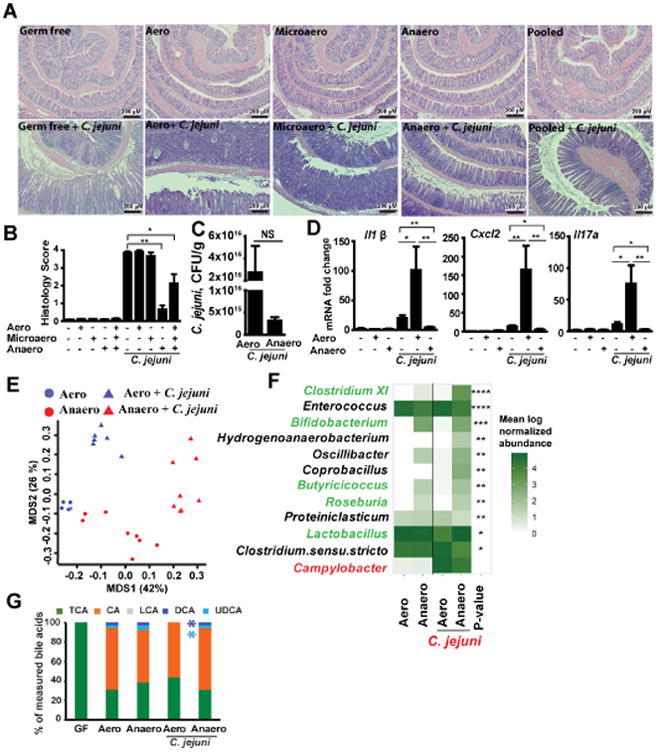
Cohorts of 4-8 GF Il10-/- mice were colonized with microbiota cultured under aerobic (Aero), microaerobic (Microaero), or anaerobic (Anaero) conditions, or all three groups pooled for 14 days. The mice were then gavaged with a single dose of 109 CFU C. jejuni/mouse and were euthanized 12 days post-infection. Stool samples from Anaero- and Aero-Biota mice were subjected to 16S rDNA sequencing and HPLC/MS analysis of bile acids. (A) H&E staining showing representative intestinal histology of C. jejuni-induced colitis in Il10-/- mice. (B) Quantification of histological intestinal damage score. (C) C. jejuni colonic luminal colonization level using culture in mice colonized with Aero- or Anaero-Biota. (D) Colonic Il1β, Cxcl2, and Il17a mRNA qPCR fold change relative to GF and normalized to Gapdh. (E) PCoA comparing Anaero- and Aero-Biota microbiome composition pre- and post-C. jejuni infection, based on 16S rDNA sequencing. (F) Heatmap representation of genera significantly different between Anaero-and Aero-Biota-colonized Il10-/- mice following C. jejuni infection, plus Campylobacter (red), which was not significant. Their abundance prior to infection is also shown. Green font indicates genera associated with anti-inflammatory response. Asterisks indicate the p-value after multiple hypothesis correction. (G) Relative stool bile acid profile measured by HPLC/MS. TCA, taurocholic acid and tauromuricholic acid; CA, cholate; LCA, lithocholic acid; UDCA, Ursodeoxycholic acid; DCA, deoxycholate. ****, P < 0.0001; ***, P < 0.001; **, P < 0.01; *, P < 0.05; NS, not significant. Scale bar is 200 μm. Results are representative of 3 independent experiments.