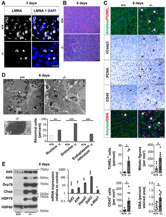
Fig. 1. Exocrine pancreas-specific Lmna-disruption triggers ER stress-response and loss of acinar cell phenotype.

(A) Loss of LMNA in most pancreatic acinar cells 3d post-tamoxifen injection. Bar=20μm. (B) Representative H&E-stained sections 6d post-tamoxifen injection. Bar=20μm. (C) Pancreata (6d) were stained as indicated; arrows highlight positive staining. Bar=50μm. Histograms: quantification using tissues from 6-9 mice/group. (D) Transmission electron microscopy of 6d control and KO-pancreata. Arrows: acinus lumen, void of secretions in KO but not control acini; arrowheads: vacuoles with electron-dense material. Bar=2μm. Lower panel: Condensed and tightly-wound ER (high magnification; bar=0.8μm). Histogram: electron-micrographs quantification (3-4 mice/group). (E) Immunoblots of indicated ER stress-related proteins, HSP70, and the mitochondrial protein HSP60. Dashed lines mark non-adjacent wells of same gel. Histogram: RT-qPCR of the indicated transcripts (N=6 mice/group). *p≤0.05,**p≤0.01,***p≤0.001.
